Association of temporomandibular joint space and condylar head position with different skeletal patterns among the Malaysian population
DOI:
https://doi.org/10.31436/ijohs.v5i2.300Keywords:
condylar head position, skeletal patternAbstract
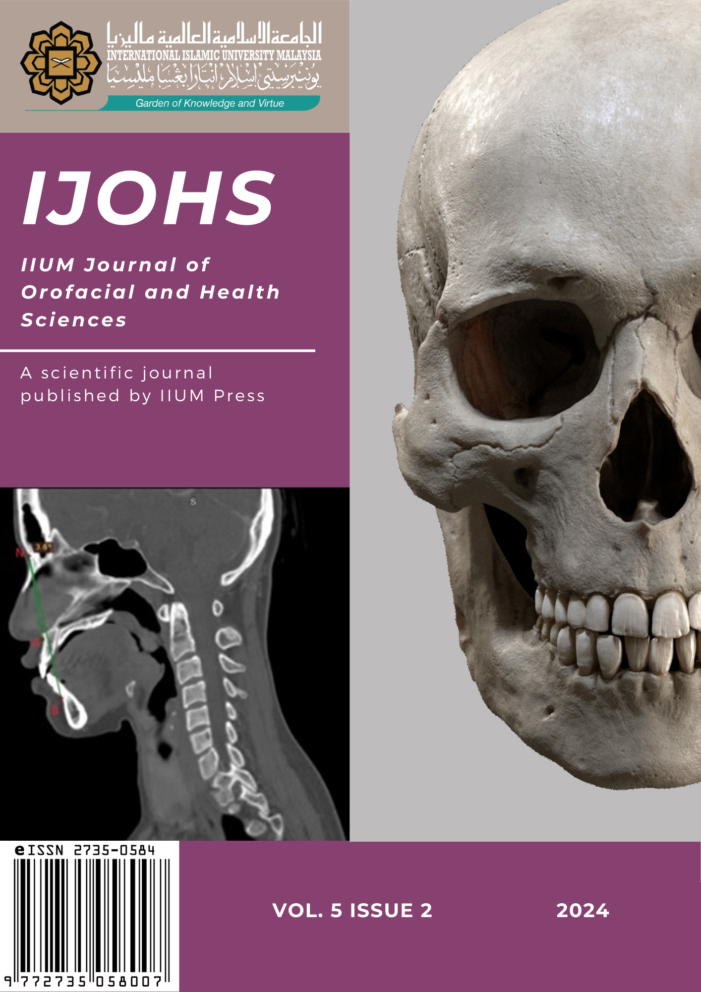
During growth and development of the head and neck, some degree of interaction and interdependence between skeletal pattern with temporomandibular joint (TMJ) space and condylar head position occurs. Results from previous studies are varied; some reporting significant difference of TMJ space or condylar head position among the skeletal patterns, whilst others have shown that no such association is present. Considering that previous studies have been conducted in populations outside of Malaysia and the importance of determining the correlation between skeletal and TMJ morphology, this retrospective study was done to evaluate the TMJ space and condylar head position in different skeletal patterns among the Malaysian population using computed tomography (CT) images. A total of 90 CT images of the head and neck were included. Skeletal pattern (class I, II, III) was determined from each CT image based on the ANB angles obtained from reconstruction of these images. The TMJ space measurement and condylar head position were determined from sagittal images based on established landmarks from the reconstructed CT images. Statistical analysis was used to compare the TMJ space and condylar head position across the three skeletal classes and assess its significance. The results of this study demonstrated that there was no significant association between TMJ space or condylar head position in the different skeletal patterns among the Malaysian population. It is recommended that a prospective study with large sample size and standardized measurement techniques be implemented in the future to determine the precise association between TMJ morphology and different skeletal patterns.
References
Alhammadi, M. S., Fayed, M. S., & Labib, A. (2016). Three-dimensional assessment of temporomandibular joints in skeletal Class I, Class II, and Class III malocclusions: Cone beam computed tomography analysis. Journal of the World Federation of Orthodontists, 5(3), 80–86. https://doi.org/10.1016/j.ejwf.2016.07.001
Chae, J.-M., Park, J. H., Tai, K., Mizutani, K., Uzuka, S., Miyashita, W., & Seo, H. Y. (2020). Evaluation of condyle-fossa relationships in adolescents with various skeletal patterns using cone-beam computed tomography. The Angle Orthodontist, 90(2), 224–232. https://doi.org/10.2319/052919-369.1
Diwakar, R., Bucci, R., Kaushik, A., Bansal, A., Bucci, P., Kochhar, A. S., & Spagnuolo, G. (2023). Three-dimensional assessment of temporomandibular joint morphology and facial asymmetry in individuals with different vertical skeletal growth patterns. International Journal of Environmental Research and Public Health, 20(2). https://doi.org/10.3390/ijerph20021437
Feres, M. F. N., Eissa, O., Roscoe, M. G., & El-Bialy, T. (2020). Comparison of the condyle sagittal position of Class I and Class II division 2 in orthodontic patients. Journal of Contemporary Dental Practice, 21(9), 977–981. https://doi.org/10.5005/jp-journals-10024-2867
Ikeda, K., & Kawamura, A. (2009). Assessment of optimal condylar position with limited cone-beam computed tomography. American Journal of Orthodontics and Dentofacial Orthopedics, 135(4), 495–501. https://doi.org/10.1016/j.ajodo.2007.05.021
Krisjane, Z., Urtane, I., Krumina, G., & Zepa, K. (2009). Three-dimensional evaluation of TMJ parameters in Class II and Class III patients. 11(1), 32–36.
Littlewood, S. J., & Mitchell, L. (2019). An Introduction to Orthodontics, 5th ed. Oxford University Press.
Liu, Z. J., King, G. J., & Herring, S. W. (2003). Alterations of morphology and microdensity in the condyle after mandibular osteodistraction in the rat. Journal of Oral and Maxillofacial Surgery, 61(8), 918–927. https://doi.org/10.1016/S0278-2391(03)00294-5
Ma, Q., Bimal, P., Mei, L., Olliver, S., Farella, M., & Li, H. (2018). Temporomandibular condylar morphology in diverse maxillary–mandibular skeletal patterns: A 3-dimensional cone-beam computed tomography study. Journal of the American Dental Association, 149(7), 589–598. https://doi.org/10.1016/j.adaj.2018.02.016
Martins, E., Silva, J. C., Pires, C. A., Ponces, M. J., & Lopes, J. D. (2015). Sagittal joint spaces of the temporomandibular joint: Systematic review and meta-analysis. Revista Portuguesa de Estomatologia, Medicina Dentaria e Cirurgia Maxilofacial, 56(2), 80–88. https://doi.org/10.1016/j.rpemd.2015.04.002
Noh, K. J., Baik, H., Han, S., Jang, W., & Choi, Y. J. (2020). Mandibular condyle and glenoid fossa morphology according to vertical and sagittal skeletal patterns: A cone beam computed tomography study. The Korean Journal of Orthodontics, 1–22.
Paknahad, M., Shahidi, S., & Abbaszade, H. (2016). Correlation between condylar position and different sagittal skeletal facial types. Journal of Orofacial Orthopedics, 77(5), 350–356. https://doi.org/10.1007/s00056-016-0039-z
Proffit, W. R., Fields, H., Larson, B., & Sarver, D. M. (2019). Contemporary Orthodontics (6e: South). Elsevier.
Pullinger, A., & Hollender, L. (1986). Variation in condyle-fossa relationships according to different methods of evaluation in tomograms. Oral Surgery, Oral Medicine, Oral Pathology, 62(6), 719–727. https://doi.org/10.1016/0030-4220(86)90270-7
Rivero-Millán, P., Barrera-Mora, J. M., Espinar-Escalona, E., Pino, C. A. G. Del, Martín-Salvador, D., & Llamas-Carreras, J. M. (2021).
Comparison of condylar position in normal occlusion, Class II Division 1, Class II Division 2 and Class III malocclusions using CBCT imaging. Journal of Clinical and Experimental Dentistry, 13(12), 1216–1226. https://doi.org/10.4317/jced.58970
Song, J., Cheng, M., Qian, Y., & Chu, F. (2020). Cone-beam CT evaluation of temporomandibular joint in permanent dentition according to Angle’s classification. Oral Radiology, 36(3), 261–266. https://doi.org/10.1007/s11282-019-00403-3
Downloads
Published
How to Cite
Issue
Section