Ossifying fibrous epulis: case reports and diagnostic insights into gingival swellings
DOI:
https://doi.org/10.31436/ijohs.v7i1.413Keywords:
gingival swelling, ossifying fibrous epulis, peripheral ossifying fibroma, reactive lesionAbstract
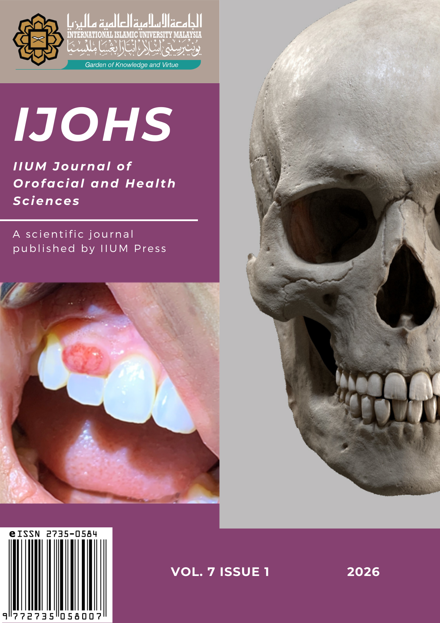
Ossifying fibrous epulis, also known as peripheral ossifying fibroma in literature, is a relatively common, benign reactive lesion that exclusively affects the gingival tissue. It arises in response to chronic local irritation such as plaque accumulation, calculus, faulty restorations, or repeated trauma. Though non-neoplastic in nature, it can present as a slowly enlarging, firm, and fibrous mass, mimicking a range of other more aggressive or neoplastic conditions. Histopathological examination is paramount for achieving a definitive diagnosis. This report presents two cases occurring in young adults, highlighting the clinical features, diagnostic process, surgical management, and post-operative outcomes. The lesions, located in the anterior maxilla and mandibular gingiva respectively, shared common clinical traits including nodular appearance, surface colour resembling surrounding mucosa, and occasional bleeding upon provocation. Histopathological analysis in both cases revealed parakeratinised stratified squamous epithelium with focal degeneration, mineralized components such as trabeculae of bone and cementum-like materials, and chronic inflammatory cells infiltrate. Following surgical excision and removal of local irritants, healing was uneventful with no recurrence observed during short-term follow-up. This paper further elaborates on the histopathological spectrum, recurrence risk, and differential diagnosis of ossifying fibrous epulis, supported by a tabulated overview of similar gingival lesions. Recognising the clinical behaviour and key distinguishing features of this lesion is crucial in general dental practice to avoid misdiagnosis and ensure appropriate intervention. Early detection, thorough removal, and patient education are integral to effective management and prevention of recurrence.
References
Agrawal, A. A. (2015). Gingival enlargements: differential diagnosis and review of literature. World Journal of Clinical Cases, 3(9), 779. DOI: https://doi.org/10.12998/wjcc.v3.i9.779
Brierley, D. J., Crane, H., & Hunter, K. D. (2019). Lumps and bumps of the gingiva: a pathological miscellany. Head and Neck Pathology, 13(1), 103–113. https://doi.org/10.1007/s12105-019-01000-w DOI: https://doi.org/10.1007/s12105-019-01000-w
Buchner, A., & Hansen, L. S. (1987). The histomorphologic spectrum of peripheral ossifying fibroma. Oral Surgery, Oral Medicine, Oral Pathology, 63(4), 452-461. DOI: https://doi.org/10.1016/0030-4220(87)90258-1
Chien, H. H., Park, J. V., Kalmar, J. R., & Javaid, S. (2024). Peripheral ossifying fibroma: a case report highlighting clinical features and management strategies. International Journal of Clinical Case Reports and Reviews, 16(5). DOI: https://doi.org/10.31579/2690-4861/376
Deepthi, T. R., Mathew, P. A., Yeshoda, T. G., Khargekar, N., Kappiamkunnath, S., & Adarsh, V. J. (2024). Peripheral ossifying ?broma: a rare case report and review of literature. Journal of Orofacial and Health Sciences, 11(2), 86-88. DOI: https://doi.org/10.18231/j.johs.2024.019
Efflom, O. A., Adeyemo, W. L., & Soyele, O. O. (2011). Focal reactive lesions of the gingiva: an analysis of 314 cases at a tertiary health institution in Nigeria. Nigerian Medical Journal, 52(1). DOI: https://doi.org/10.4103/0300-1652.80074
Gulati, R., Khetarpal, S., Ratre, M. S., & Solanki, M. (2019). Management of massive peripheral ossifying fibroma using diode laser. Journal of Indian Society of Periodontology, 23(2), 177–180. https://doi.org/10.4103/jisp.jisp_431_18 DOI: https://doi.org/10.4103/jisp.jisp_431_18
Katanec, T., Budak, L., Brajdi?, D., & Gabri?, D. (2022). Atypical peripheral ossifying fibroma of the mandible. Dentistry Journal, 10(1), 9. https://doi.org/10.3390/dj10010009 DOI: https://doi.org/10.3390/dj10010009
Krishna, V.K, Periasamy, S., Kumar, S.P., Bhat, S.V. (2022). Atypical presentation of peripheral ossifying fibroma in the mandible. Cureus, 14(2), e22375. https://doi.org/10.7759/cureus.22375 DOI: https://doi.org/10.7759/cureus.22375
Maturana-Ramírez, A., Adorno-Farías, D., Reyes-Rojas, M., Farías-Vergara, M., & Aitken-Saavedra, J. (2015). A retrospective analysis of reactive hyperplastic lesions of the oral cavity: study of 1149 cases diagnosed between 2000 and 2011, Chile. Acta Odontológica Latinoamericana, 28(2), 103-107.
Sacks, H. G., Amrani, S., & Anderson, K. (2012). “Gigantiform” peripheral ossifying fibroma: report of a case. Journal of Oral and Maxillofacial Surgery, 70(11), 2610-2613. DOI: https://doi.org/10.1016/j.joms.2011.12.011
Takagi, R., Mori, K., Koike, T., Tsuyuguchi, S., Kanai, K., Watanabe, Y., et al. (2024). A giant peripheral ossifying fibroma of the maxilla with extreme difficulty in clinical differentiation from malignancy: a case report and review of the literature. Journal of Medical Case Reports, 18(1). https://doi.org/10.1186/s13256-024-04529-9 DOI: https://doi.org/10.1186/s13256-024-04529-9
Zhao, N., Yesibulati, Y., Xiayizhati, P., He, Y. N., Xia, R. H., & Yan, X. Z. (2023). A large-cohort study of 2971 cases of epulis: focusing on risk factors associated with recurrence. BMC Oral Health, 23(1), 229. https://doi.org/10.1186/s12903-023-02935-x DOI: https://doi.org/10.1186/s12903-023-02935-x
Downloads
Published
How to Cite
Issue
Section