SUBMISSION OF CASE REPORTS
Important notice to all authors

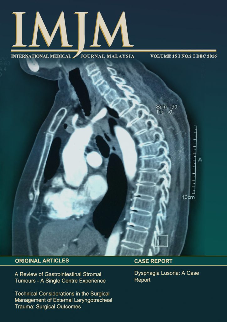
Sagittal view CT scan picture showing aberrant right subclavian artery compressing on the oesophagus.
IMJM Vol 15 Supplement
Editorial Committee
Pure Science
Prof. Dr. Ahmed Jalal Khan Chowdhury
Clinical Medicine
Assoc. Prof. Dato’ Dr. Mohd Basri Mat Noor
Assoc. Prof. Dr. Mohamed Saufi Awang
Public Health
Assoc. Prof. Dr. Jamalludin Ab. Rahman
Asst. Prof. Dr. Norazlina A. Rahman
Basic Health Sciences
Prof. Dr. Naznin Muhammad
Assoc. Prof. Dr. Farahidah Mohamed
IIUM Medical Centre – A Reality
A state-of-the-art 300 bedded Teaching hospital situated in Kuantan Campus scheduled to open in July 2016

Aedes aegypti, a primary vector for dengue feeding on human blood
Magnified picture of a fine-needle aspiration smear in Papanicolaou staining with cytologic features mistaken for carcinoma, including sheets of cells with pleomorphic, hyperchromatic nuclei, prominent nucleolus and moderate cytoplasm.
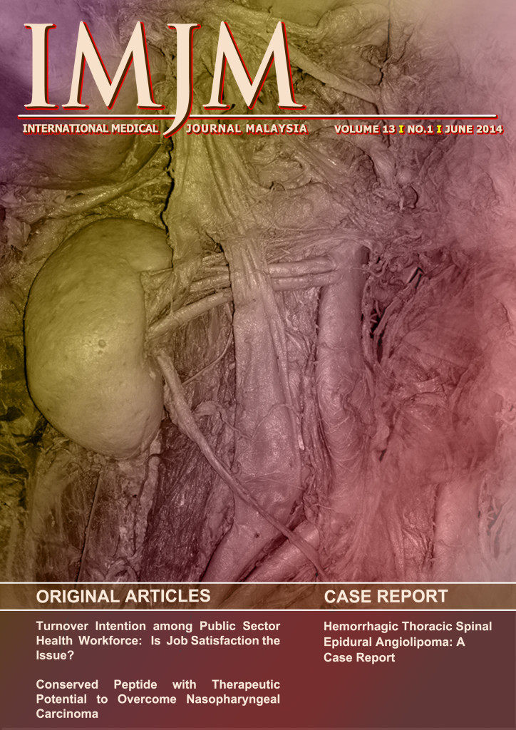
Picture shows an abdominal dissection of a 65 years old male cadaver, with multiple variations of the right kidney vasculature. There were double right renal arteries with prehilar branching of the upper renal artery. The renal vein ascended upwards obliquely before ending in the lateral aspect of the inferior vena cava.
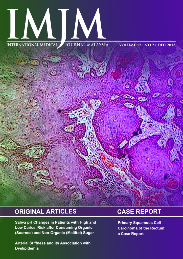
‘Picture from a histopathological specimen of a well-differentiated squamous-cell carcinoma arising from the rectal mucosa'
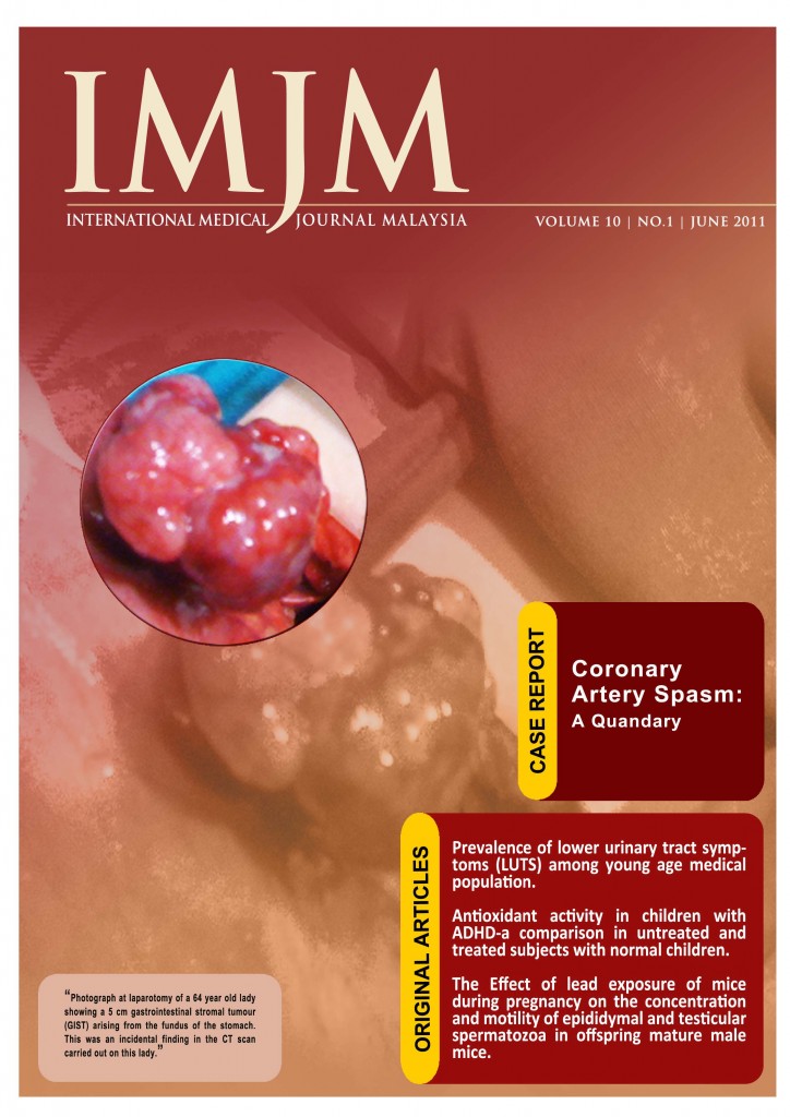
Photograph at laparotomy of a 64 year old lady showing a 5 cm gastrointestinal stromal tumour (GIST) arising from the fundus of the stomach. This was an incidental finding in the CT scan carried out on this lady.
![]()
![]()
![]()
![]()
![]()
![]()
![]()

Important notice to all authors
